
Comparing Image Reconstruction Methods with Electrical Impedance Tomography Data
Electrical impedance tomography (EIT) is a noninvasive imaging technique that uses electrodes to detect electrical conductivity of biological tissue and reconstruct tomographic images of the human body. The electrodes apply currents to specific bodily regions and measure the voltage; practitioners then utilize the resulting data to solve an inverse problem and reconstruct the conductivity and permittivity of the area in question (see Figure 1). EIT is a popular approach because it is portable, noninvasive, and low cost compared to other medical imaging methods; avoids ionizing radiation; and allows for continuous monitoring in real time. As such, it is frequently used to examine breast tissue and monitor lung activity, epileptic seizures, and strokes. Beyond the medical field, EIT can assess the structural characterization of soil and rock and discern the health of civil infrastructure, such as bridges and support beams.
During the 2025 SIAM Conference on Computational Science and Engineering, which took place last week in Fort Worth, Texas, Jacob Roarty of Arizona State University employed EIT data to compare the effectiveness of various image reconstruction techniques—including iterative, noniterative, and machine learning processes—that identify and locate anomalies in the human body. He began with a forward problem, which involves a series of partial differential equations that account for the conductivity, voltage, boundary, outward unit normal vector, surface area of the electrode, and injected current. These equations also establish a ground truth and account for contact impedance: a highly resistant region between the electrode and body. Using simulated data, Roarty established a forward model with 16 electrodes and 2,959 mesh elements. He used a semi-uniform mesh, which easily detects incongruities that are close to the electrodes but struggles to sense those that are further away.
![<strong>Figure 1.</strong> Overview of electrical impedance tomography (EIT) and the resulting image generation and interpretation process. Figure courtesy of [1].](/media/ynblxm14/figure1.jpg)
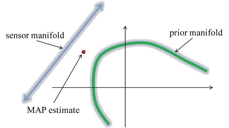
In practice, one can represent the EIT problem via a discrete model with a defined sensitivity matrix. However, the singular value decomposition (SVD)—which yields 104 unique measurements on a 16-electrode system—confirms that the inverse of this problem is severely ill-posed. Limited measurements from the partial boundary data—coupled with electrical properties within the body that cause regularization difficulties—are responsible for this ill-posedness, though a truncated SVD (TSVD) somewhat mitigates the issue. “Given the ground truth and our reconstruction, we do a fairly decent job,” Roarty said. “We get a rough idea of where the anomaly is and a good idea of the conductivity, but we also get a little bit of noise.” Tikhonov regularization produced a similar result.
Acknowledging the limitations of these types of conventional noniterative mechanisms, Roarty turned to other approaches. He next iterated via the Gauss-Newton method, which follows a similar methodology as the Tikhonov regularization. When utilizing this method, one must choose an initial conductivity approximation, solve a forward problem to obtain simulated measures, and update conductivity to ultimately yield a new sensitivity matrix, repeating as necessary. “Rather than a blanket guess, we now have an informed guess that we hope will give us better results,” Roarty said.
For the purposes of comparison, Roarty also experimented with another iterative process called total variation (TV). “It’s really a primal dual-interior point method with a TV component,” he said. While Gauss-Newton certainly showed some improvement over noniterative methods, TV eradicated the noise after 10 iterations and provided better edge preservation.
Because noniterative methods struggle with sharp boundaries and iterative methods are both expensive and time consuming, Roarty concluded his presentation with a brief exploration of deep neural networks (DNNs). Specifically, he investigated the effectiveness of a “hemiarray” (i.e., half array) model, which places eight electrodes—rather than the full set of 16—on the torso. This type of setup is especially effective for patients whose conditions could be exacerbated by lifting/shifting them to apply the full array, such as individuals with abdominal trauma or premature infants (see Figure 2).
![<strong>Figure 2.</strong> Deep neural networks make it possible for practitioners to utilize a hemiarray of eight electrodes—rather than the full set of 16—to detect anomalies in medically fragile patients, such as premature infants or those with abdominal trauma. Figure courtesy of [1].](/media/v23c25cl/figure2.jpg)
While traditional methods that only use eight electrodes would lose too much data to garner meaningful results, Roarty was able to feed the hemiarray inputs through a neural network and retain the full range of outputs from the 16-electrode framework. Thus, the DNN method can successfully identify anomalies even when they are not within the immediate vicinity of the electrodes. “This is where the neural network assists us in retrieving some of the missing data,” Roarty said.
Jacob Roarty received funding to attend CSE25 through a SIAM Student Travel Award. To learn more about Student Travel Awards and submit an application, visit the online page.
SIAM Student Travel Awards are made possible in part by the generous support of our community. To make a gift to the Student Travel Fund, visit the SIAM website.
References
[1] Adler, A., & Holder, D. (Eds.). (2022). Electrical impedance tomography: Methods, history, and applications (2nd ed.). In Series in medical physics and biomedical engineering. Boca Raton, FL: CRC Press.
About the Author
Lina Sorg
Managing editor, SIAM News
Lina Sorg is the managing editor of SIAM News.

Related Reading

Stay Up-to-Date with Email Alerts
Sign up for our monthly newsletter and emails about other topics of your choosing.
