Exploring the Radon Transform and the Field of Medical Imaging
The following is a brief reflection from the author of The Radon Transform and Medical Imaging, which was published by SIAM in 2013 as part of the CBMS-NSF Regional Conference Series in Applied Mathematics. The text surveys the main mathematical concepts and techniques that drive both well-established imaging modalities and developing methodologies. It explores a variety of concepts that pertain to medical imaging, including stability, inversion, incomplete data effects, and the role of interior information.
The Radon Transform and Medical Imaging addresses the topics of 10 lectures that I delivered during the 2012 NSF-CBMS Conference on Mathematical Methods of Computed Tomography. Tomography aims to produce a picture of the internal slices of a nontransparent body, such as a patient’s physique, an airplane wing, or the Earth itself. It does so by sending signals—ultrasound, X-rays, electrical currents, light, and so forth—to penetrate the body in question, then recording the outcome. In other words, the body acts like a “black box” into which practitioners send signals and examine the resulting outputs. We must then recover the hidden mechanism from these observations.
However, we cannot address the problem with “black box” generality. Signal propagation (and thus observable output) is governed by a “hidden” partial differential equation (PDE). Although we usually possess some general knowledge about the equation, we are often not sure of its coefficients. The coefficients therefore comprise the overall picture that we seek. This setup is called an inverse problem; we know the answers but not the questions that are hidden inside the box. One famous example of such a problem is evident in Douglas Adams’ The Hitchhiker’s Guide to the Galaxy, which discusses the “ultimate question of life, the universe, and everything.” The answer, calculated by a supercomputer in a matter of 7.5 million years, was 42. But no one knew what the question was!
The wide range of important applications for computed tomography (CT) explains why it has been attracting the attention of medical practitioners, mathematicians, physicists, engineers, geophysicists, and other scientists for the last half-century. In addition to its usefulness, CT boasts a whole world of mathematical techniques and problems that delight both pure and applied mathematicians: commutative and non-commutative Fourier (harmonic) analysis; differential equations; geometry (integral, differential, and algebraic); complex analysis (in several variables); microlocal analysis; group representation theory; discrete mathematics; probability theory and statistics; and numerical analysis.
The audience of the 2012 NSF-CBMS conference encompassed a wide range of attendees, from graduate students to established researchers. I therefore attempted to keep the exposition as self-contained as possible; in most cases, experts should skip the background information in the appendices to avoid feeling patronized. I also tried to minimize technicalities and instead emphasize the main mathematical ideas and intuitions. Readers can develop the details to their full extent or find them in the literature (the text contains pointers to help them do so) — this framework explains the book’s rather colloquial style. Here I briefly describe the contents of The Radon Transform and Medical Imaging.
The first part of the text introduces readers to medical imaging and a variety of relevant problems, including image reconstruction (the goal of the book), processing, and interpretation. It then explains the notion of CT and provides a classification into transmission, emission, and reflection types. I also present various application areas beyond medicine, such as industry; homeland security; geology, geophysics, and seismology; radar, sonar, and lidar; and even archeology.
The text then lists the milestones in the subject’s more than 100-year history and discusses contrast, stability, and resolution. Since nearly all relevant methods involve the recovery of a known PDE’s unknown coefficients, the early chapters address the types of equations that arise in several common CT techniques. Knowledge of the type of equation (hyperbolic, parabolic, elliptic, etc.) offers users important information about the kind of resolution they can expect.
The book’s second part overviews the basic mathematics of the most familiar traditional techniques: X-ray CT and emission tomography. Chapters five and six tackle topics like inversion, reconstruction stability, and artifacts and their microlocal origins. This segment proceeds to present the main notions of CT (such as backprojection) and derives important formulas (e.g., inversion formulas, the Fourier slice theorem, and range conditions).
Chapter seven examines the important issue of artifacts in reconstruction and the microlocal approaches to understand and predict them. While learning microlocal analysis in its full capacity is a daunting task for a beginner, microlocal analysis results and tests are very easy to use. Chapter nine then briefly surveys numerical techniques, which could fill a book by themselves, and chapter 10 briefly describes other popular medical imaging modalities like magnetic resonance imaging, optical tomography, and ultrasound.
The third part of the text introduces an abundance of newly developed, so-called hybrid techniques. Since each known modality suffers from some drawbacks (e.g., low resolution or low contrast), the hybrid methods strive to combine them to alleviate their individual faults and capitalize on their advantages. The main concentration here is thermo-(opto-)acoustic tomography, ultrasound modulation of electrical and optical tomography, and problems with “internal information.”
Finally, the book’s fourth and final part consists of appendices that address the harmonic analysis, differential equations, and functional analysis tools that I utilize throughout the text.
The Radon Transform and Medical Imaging does not assume that readers are familiar with CT, which makes it accessible to a wide audience of specialists and nonexperts alike. Graduate students and researchers in mathematics, engineering, and physics who wish to learn more about medical imaging will all benefit from reading this text.
Enjoy this passage? Visit the SIAM Bookstore to learn more about The Radon Transform and Medical Imaging and browse other SIAM titles.
About the Author
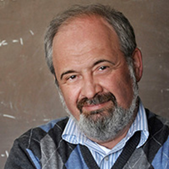
Peter Kuchment
Distinguished Professor, Texas A&M University
Peter Kuchment is a University Distinguished Professor of Mathematics at Texas A&M University. His research areas of interest include spectral theory, partial differential equations, mathematical physics, inverse problems, and imaging. Kuchment is also a SIAM Fellow.

Stay Up-to-Date with Email Alerts
Sign up for our monthly newsletter and emails about other topics of your choosing.


